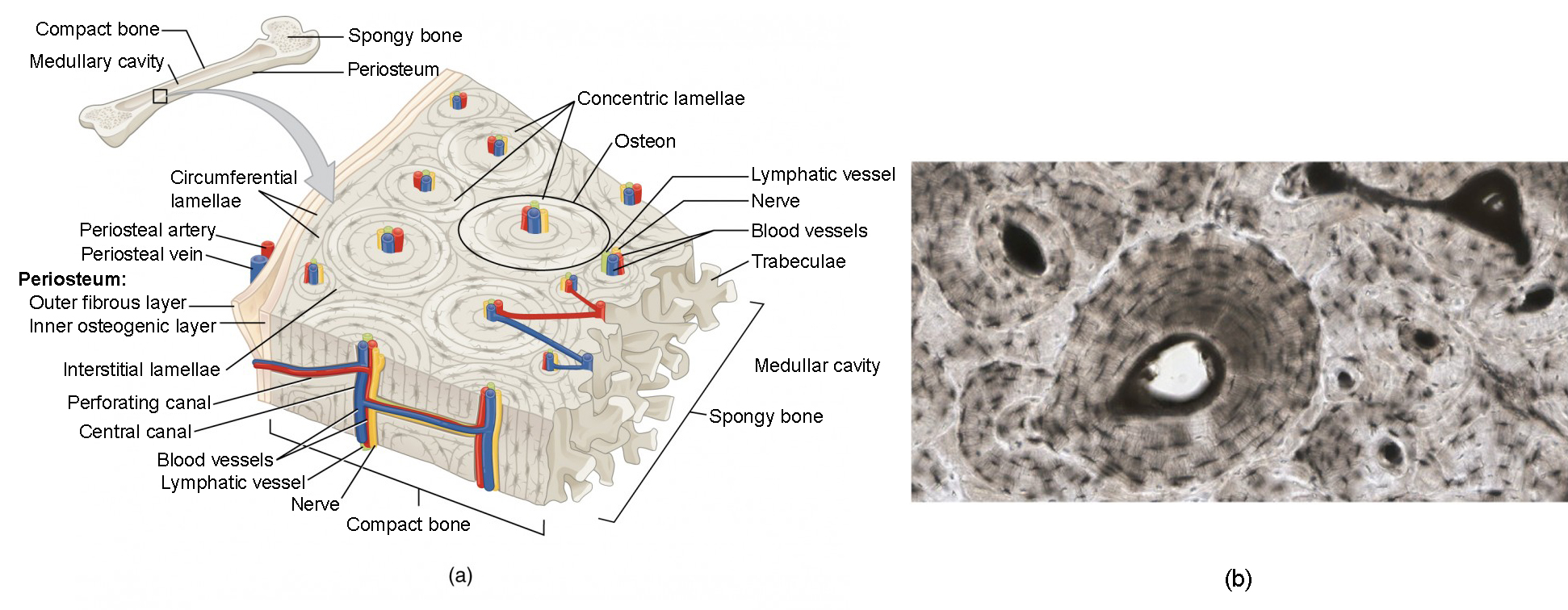
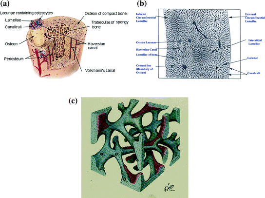
Compact Bone Diagram Unlabeled / Skeleton Worksheet Wikieducator : The osteon consists of a central canal called the osteonic (haversian) canal, which is surrounded by concentric rings (lamellae) of matrix.
Compact Bone Diagram Unlabeled / Skeleton Worksheet Wikieducator : The osteon consists of a central canal called the osteonic (haversian) canal, which is surrounded by concentric rings (lamellae) of matrix.. This lab is designed to provide students with an overview of bones through a variety of investigative to identify the major regions and structures of an osteon in a histological specimen of compact bone (or diagram or model of one). Muscles leg part posterior compartment. Sclerostin inhibits bone formation mostly by antagonizing lrp5/6, thus inhibiting wnt signaling. A typical long bone showing gross anatomical features. 6 compact bone vs spongy bone.
(micrograph provided by the regents of university of michigan. Its unlabeled, so that your practce better. Hand, grasping organ at the end of the forelimb of certain vertebrates that exhibits great mobility and flexibility in the digits and in the whole organ. Students fill in the boxes with the names of the bones. Compact bone consists of closely packed osteons or haversian systems.
Human skeleton diagram unlabeled graph diagram.
Bone classification, structure & relationships: Compact bone consists of closely packed osteons or haversian systems. The outer part of a long bone is made of compact bone. Long bone structure diagram and definitions flashcards quizlet. Sclerostin inhibits bone formation mostly by antagonizing lrp5/6, thus inhibiting wnt signaling. Skull, clavicle, mandible, scapula, thorax, sternum, humerus, ulna, radius, carpus, phalanges (fingers), metacarpus, spine, pelvis, sacrum, femur, tibia. Appendicular skeleton quiz diagram clip art at clker com vector. Hand, grasping organ at the end of the forelimb of certain vertebrates that exhibits great mobility and flexibility in the digits and in the whole organ. Human tongue anatomy vector image. Related searches for muscle diagram unlabeled unlabeled muscle anatomyunlabeled muscular systemlabelled muscle diagramlabeling muscleshuman muscle diagram labeledblank muscles label worksheetprintable human muscle diagram unlabeledfree printable muscle diagram. What are diplo , its function, and location? The osteon consists of a central canal called the osteonic (haversian) canal, which is surrounded by concentric rings (lamellae) of matrix. Human skeleton diagram unlabeled graph diagram.
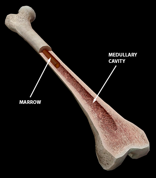
Femur bone diagram unlabeled via. Long bone structure diagram and definitions flashcards quizlet. 6 compact bone vs spongy bone. Students fill in the boxes with the names of the bones. Location of red and yellow marrow in adults and.
Related images with foot bone diagram unlabled.
Anchor chart human bone diagram human body skeleton stem science health hand. Bone classification, structure & relationships: Total there are 12 pairs of ribs, as you can see in the diagram. What are diplo , its function, and location? Key.' carotid canal coronal suture ethmoid bone external occipital protuberance foramen lacerum foramen magnum foramen ovale frontal bone edwnq'p'iep'n glabella. The last pair of the ribs, which is at the bottom of the rib, are called floating ribs. Sclerostin inhibits bone formation mostly by antagonizing lrp5/6, thus inhibiting wnt signaling. Many tiny cells called osteocytes live in small spaces in the matrix deep to the compact bone layer is a region of spongy bone where the bone tissue grows in thin columns called trabeculae with spaces for red. Learn vocabulary, terms and more with flashcards, games and other study tools. Appendicular skeleton quiz diagram clip art at clker com vector. Femur bone diagram unlabeled via. (b) in this micrograph of the osteon, you can clearly see the concentric lamellae and central canals. Practice quiz & test prep for students and teachers.
Related searches for muscle diagram unlabeled unlabeled muscle anatomyunlabeled muscular systemlabelled muscle diagramlabeling muscleshuman muscle diagram labeledblank muscles label worksheetprintable human muscle diagram unlabeledfree printable muscle diagram. The bones mentioned in each human skeleton chart are: Many tiny cells called osteocytes live in small spaces in the matrix deep to the compact bone layer is a region of spongy bone where the bone tissue grows in thin columns called trabeculae with spaces for red. This simple worksheet shows a skeleton with bones unlabeled. Compact bone is made of a matrix of hard mineral salts reinforced with tough collagen fibers.
Location of red and yellow marrow in adults and.
The bones mentioned in each human skeleton chart are: Compact bone forms the outer layer of all bones and most of the structure of long bones see diagram right. Key.' carotid canal coronal suture ethmoid bone external occipital protuberance foramen lacerum foramen magnum foramen ovale frontal bone edwnq'p'iep'n glabella. (micrograph provided by the regents of university of michigan. Sclerostin inhibits bone formation mostly by antagonizing lrp5/6, thus inhibiting wnt signaling. Location of red and yellow marrow in adults and. Long bone structure diagram and definitions flashcards quizlet. Human gross anatomy study | humandiagram.info. .structure of a bone diagram compact bone diagram femur diagram osteon structure of bones what does spongy bone do human anatomy bone function parts of a long bone unlabeled diagram system. Structure of compact bone longitudinal and cross sectional view of download scientific diagram. Label compact and spongy bone illustrations as demonstrated in class. The bones shown in the chest and hip region in the labeled human skeleton diagram are the ribs, vertebrae, pelvis, os coxae, sacrum and coccyx. The osteon consists of a central canal called the osteonic (haversian) canal, which is surrounded by concentric rings (lamellae) of matrix.
Related searches for muscle diagram unlabeled unlabeled muscle anatomyunlabeled muscular systemlabelled muscle diagramlabeling muscleshuman muscle diagram labeledblank muscles label worksheetprintable human muscle diagram unlabeledfree printable muscle diagram compact bone diagram. Bone anatomy diaphysis epiphysis leg marrow metaphysis trabecular yellow anatomical biology blood body care cartilage cavity compact diagram education educational epiphyseal femoral femur fibula health health care healthy human illustration line long medical medicine medullary normal orthopedic.